CYSTS OF JAW

INTRODUCTION
A cyst is a pathological cavity lined by epithelium containing fluid or semi-fluid contents.
Cysts are common in the jaw bones as these bones contain epithelium left after tooth development.
Cysts of the jaw can be classified into two main groups: odontogenic cysts (cysts lined by odontogenic epithelium, i.e. epithelium derived from the dental lamina) and non-odontogenic cysts (cysts lined by other types of epithelium).
The classification of common jaw cysts.
| Odontogenic cysts | Non-odontogenic cysts |
|
|
Radicular cysts
$ads={2}
Radicular cysts are the most common type of jaw cyst. They are always found in association with a non-vital tooth.
Radicular can be classified depending upon their location in relation to the tooth:
- Periapical: found at the apex
- Lateral: found alongside the tooth root
- Residual: persists in the tooth extraction socket following tooth removal
Aetiology
Many non-vital teeth exist without associated radicular cysts, however, inflammation can induce cystic development.
The cyst initially develops following the proliferation of Rests of Malassez. These are small epithelial remnants of Hertwig’s epithelial root sheath, an important feature in tooth root development. As these cells proliferate and grow a central cavity is formed from the autolyses of central cells. This central cavity contains tissue fluid and cellular debris which produces hydrostatic pressure leading to cyst enlargement.
Clinical features
Initially, when the radicular cyst is small it is unlikely to have any presenting features. There is likely to be a history of toothache from the causative tooth though this may precede cyst development by many years.
As the cyst grows there may be a noticeable swelling in the buccal sulcus adjacent to the causative tooth. This swelling is often described as having ‘eggshell’ like features meaning if it is pressed a cracking sensation may be felt. If the swelling is particularly large tooth displacement and/or mobility may be noticed.
If the cyst becomes infected, it may become painful.
Investigations
Radiographic features
A radicular cyst typically presents as a unilocular, rounded radiolucency with a well-defined outline found most often around the apex of a tooth or along its side. If it is a large cyst bucco-lingual expansion may be noted. Root resorption is rare.
Histology
Unless the cyst is small and the diagnosis is certain from clinical findings, in a hospital setting, cysts are commonly sent for histopathological analysis following their removal.
A radicular cyst wall contains fibrous connective tissue with markers of inflammation such as plasma cells. The wall also contains cholesterol clefts and hemosiderin (a breakdown product of blood cells). The central lumen is surrounded by a non-keratinised epithelium of varying thickness.
Management
Dental extraction is typically required, particularly as radicular cysts tend to develop in poor dental attendees with grossly carious, unrestorable teeth.
There is some debate amongst the literature with regard to the success of endodontic treatment for radicular cysts. This is a particularly difficult area to study in humans where the definitive diagnosis of a radicular cyst can only be confirmed histologically. It is generally accepted however that should the patient be willing (and informed of the risks of further treatment) endodontic treatment is a good first course of treatment.
Following extraction/endodontic treatment the cyst should be reviewed radiographically for signs of shrinkage. If the cyst persists, further surgical treatment such as enucleation may be required.
Dentigerous cyst
Dentigerous cysts are always found around the crown of an unerupted tooth, attaching to the cemento-enamel junction.
Aetiology
Whilst they are considered developmental, inflammation (e.g. from pericoronitis) may initiate them. Pressure on the tooth follicle from an impacted tooth leads to a reduction in venous drainage and fluid accumulation between the reduced enamel epithelium and the enamel of the tooth. The cyst then enlarges by internal pressure expanding the dental follicle.
Clinical features
Dentigerous cysts are typically seen in younger patients, aged 20-50 years old.
They are often asymptomatic and found incidentally on radiographs. Dentigerous cysts are most commonly found around the teeth which are more likely to become impacted (lower 8’s, upper 3’s). With continued growth bony expansion may be noted. As with all cysts, if they become infected, they may be painful.
Investigations
Radiographic features
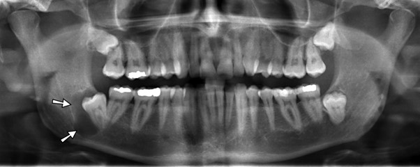
Dentigerous cysts present as a well-defined, unilocular radiolucency found around the crown of an unerupted tooth. Tooth displacement is common and root resorption of adjacent teeth is known to occur. Figure 1 shows an example dentigerous cyst.
Histopathology
Following removal of a suspected dentigerous cyst, it is always sent for histopathological analysis. This is particularly important for cysts around the angle of the mandible where the differential diagnosis includes odontogenic keratocysts (which will be discussed next) and ameloblastoma.
A dentigerous cyst will have a thin, non-keratinised squamous cell epithelium that is continuous with the reduced enamel epithelium of the tooth.
Management
Management of dentigerous cysts usually involves extraction of the causative tooth and cyst enucleation. If a clinical need exists to preserve the tooth, marsupialisation can be attempted. If a cyst is large, around a lower 8, the patient must be warned about the possibility of iatrogenically fracturing the mandible during its removal.
Odontogenic keratocyst
Odontogenic keratocysts make up about 5-10% of jaw cysts and have distinctive clinical, radiographic and histological features.
Aetiology
Odontogenic keratocysts arise from the rests of Serres (a rest of odontogenic epithelium which remains after tooth formation). Many are linked to a mutation of the PTCH gene.
Clinical features
Odontogenic keratocysts are usually found in the mandible, often around the angle. Like other cysts, it is often asymptomatic until large or infected.
Swelling is possible, though less common with odontogenic keratocysts as the cyst will spread through the medullary cavity of the bone (ie in a proximal-distal direction) before expanding in a bucco-lingual direction. Therefore, patients with bucco-lingual swelling often have a large underlying cyst.
If the cyst contents are aspirated a white fluid is seen- indicative of keratin.
Investigations
Radiographic features
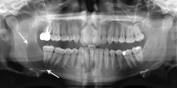
Odontogenic keratocysts present as a well-defined radiolucency. This is often multilocular though can also be unilocular. As previously mentioned, there is often evidence of growth along the medulla of the bone with little cortical expansion (Figure 2).
Histopathology
As previously discussed, all cysts presenting as a radiolucency around the angle of the mandible are sent for histopathological analysis.
An odontogenic keratocyst has distinctive histopathological features including:
- Uniform thickness, thin epithelial layer
- Satellite cysts within the wall (important as these can lead to recurrence)
- The lining epithelium is corrugated with a thin parakeratinsed surface
Management
Enucleation of the odontogenic keratocyst is the mainstay of treatment.
However, unlike other cysts, if treated by this alone the odontogenic keratocyst will usually recur. This is due to the presence of Satellite cysts with the cyst wall which itself is thin and friable and likely to break during removal. To overcome this, surgeons will typically use an intra-operative fixative (such as Carnoys Solution) to degenerate the remaining tissue.
Lateral periodontal cyst
Lateral periodontal cysts are uncommon however there is a tendency amongst dental students to misdiagnose lateral radicular cysts as them. They are found as unilocular, round radiolucency’s on the lateral surface of tooth roots (typically a lower canine/premolar).
Lateral periodontal cysts can be distinguished from a radicular cyst as often the associated tooth is vital.
These cysts should be enucleated and sent to histopathology however if small, asymptomatic and the associated tooth is proven to be vital it can be left and closely monitored.
Nasopalatine duct cyst
A nasopalatine duct cyst is a non-odontogenic cyst, which is sometimes called an incisive canal cyst. These typically present in 30-60-year-olds.
Aetiology
A nasopalatine duct cyst is thought to be derived from epithelial residues in the nasopalatine canal.
Clinical features
Nasopalatine duct cysts typically present as a swelling in the anterior, midline palate. There may be associated pain/discharge or tooth mobility/displacement if particularly large. The anterior teeth are vital.
Investigations
Radiological features
When nasopalatine duct cysts are small on radiographs it can sometimes be difficult to distinguish them from the normal appearance of a large incisive foramen.
When nasopalatine duct cysts are large, they present as a round, unilocular, corticated uniform radiolucency. They can appear heart-shaped either due to notching by the nasal septum or superimposition of the nasal spine.
Histopathology
Histopathological findings of nasopalatine duct cysts can be very variable.
Typically, respiratory epithelium that is pseudo-stratified and ciliated is seen but it can also be stratified squamous, columnar or cuboidal. Specialised respiratory cells such as goblet cells are occasionally found. Neurovascular bundles are found in most nasopalatine cyst walls.
Management
As with other cysts, the mainstay of management of nasopalatine duct cysts is surgical enucleation.
Nasolabial cyst
Nasolabial cysts are also known as a nasoalveolar cyst. These are exceedingly rare development cysts with a limited number of reports. Due to this, there is a wide age distribution in the literature (from 12 to 75 years).
Aetiology
Due to the limited number of nasolabial cyst cases, precise aetiology is difficult to define. There is however consensus that the cyst seems to develop from epithelial remnants of the nasolacrimal duct/rod.
Clinical features
Swelling is often the only clinical feature, typically of the lip and nasolabial fold. If particularly large there may be a degree of nasal obstruction.
Investigations
Radiological features
A nasolabial cyst presents as a radiolucency above the apices of the incisor teeth. If large there may be resorption of the inferior nasal notch.
Histopathology
A nasolabial cyst has a non-ciliated pseudostratified columnar epithelium which may include goblet cells. The cyst wall is relatively acellular. Nasolabial cysts are extra-osseous but lie subperiosteally.
Management
Careful surgical enucleation is the primary treatment for nasolabial cysts.
Solitary bone cyst
Solitary bone cysts are rare fluid/gas-filled lesions typically found in the metaphyseal region of long bones in young patients though can also present in the jaw.
Clinical features
Solitary bone cysts are more common in the mandible and are often seen in the premolar region. Almost all maxillary cases are found anteriorly.
Solitary bone cysts are typically asymptomatic and seen as an incidental finding on radiographs. If particularly large they are known to cause swelling, pain or paraesthesia.
Investigations
Radiological features
Solitary bone cysts present as a well-defined, unilocular, corticated radiolucency with often little cortical expansion. Scalloping between the roots of the teeth is common.
Histopathology
Solitary bone cysts are often found to be empty. They may consist of loose fibrous tissue with no epithelial lining.
Management
Surgical management of solitary bone cysts is usually only done to aid diagnosis. The cyst wall is curreted and this results in healing in most cases.
Aneurysmal bone cyst
Aneurysmal bone cysts are uncommon, blood-filled cysts which can be found in any bone though the majority are found within the long bones/spine.
Clinical features
In the jaw, aneurysmal bone cysts present as a firm swelling which is often not painful. This progressively worsens at a rate described as reasonably rapid. Aneurysmal bone cysts are often found at the angle of the mandible and depending upon their size may cause trismus.
Investigations
Radiological features
An aneurysmal bone cyst presents as a multilocular radiolucency with cortical expansion. It can be difficult to differentiate clinically and radiographically from other similar lesions (including odontogenic keratocyst and ameloblastoma).
Histopathology
As the name “aneurysmal bone cysts” suggests the lesion consists of many blood vessels. There is typically cellular fibrous tissue containing multiple blood lakes. Small multinucleate cells (giant cells) and osteoid/woven bone are also commonly found.
Management
It is first important to distinguish the aneurysmal bone cyst from other lesions-typically by taking an aspirate which will demonstrate blood. This is vital as approaching the cyst surgically without this knowledge could lead to a catastrophic bleed.
The most frequent treatment of an aneurysmal bone cyst is curettage of the lesion. There is a need for close follow up as the recurrence rates are high.
Summary table
A summary of the three most encountered cysts of the jaw.
| Radicular cyst | Dentigerous cyst | Odontogenic keratocyst |
Most likely location | Associated with a non-vital tooth (typically the apex) | Around the crown of an unerupted tooth (typically a lower 8) | Angle of the mandible |
Clinical features | Often preceding toothache The cyst can be asymptomatic if small Can have facial swelling/pain if large or infected | Often asymptomatic if small If infected can get pain/facial swelling | Often asymptomatic initially. Spreads through the medullary cavity first so bucco-lingual expansion of the mandible is often a late sign |
Radiographic features | Well defined, unilocular radiolucency associated with a non vital tooth | Well defined, unilocular radiolucency around the crown of an unerupted tooth | Well defined, multilocular (though can be unilocular) radiolucency around the angle of the mandible |
Histopathological features | Cyst wall contains fibrous connective tissue, cholesterol clefts and hemosiderin The central lumen is surrounded by a non-keratinised epithelium of varying thickness | Thin, non-keratinised squamous cell epithelium that is continuous with the reduced enamel epithelium of the tooth | Uniform thickness, thin epithelial layer Satellite cysts within the wall The lining epithelium is corrugated with a thin parakeratinsed surface
|
Management | Extraction of causative tooth (or endodontic treatment of tooth if clinically appropriate) Enucleation of cyst if large/persistent after extraction/ endodontics | Enucleation of the cyst with extraction of the associated tooth | Enucleation of the cyst followed by use of a fixative (Carnoy’s solution) |
Key points
- A cyst is a pathological cavity lined by epithelium and filled with fluid.
- The bones of the jaw contain odontogenic epithelium. Therefore cysts are more commonly found in the jaw than any other bone of the body.
- Cysts of the jaw can be categorised according to their epithelium as either odontogenic or non-odontogenic.
- Jaw cysts have a variable clinical presentation ranging from asymptomatic to jaw pain/facial swelling.
- A radicular cyst is by far the most common jaw cyst. These are always associated with a non-vital tooth.
- There are numerous possibilities for a cyst presenting as a radiolucency at the angle of the mandible which include dentigerous cyst and odontogenic keratocyst.
- Due to the often overlapping clinical and radiographic features, the majority of cysts are sent to histopathology following removal.
- Aspirating a cyst prior to removing it may give an indication of the diagnosis. This is especially important when an aneurysmal bone cyst is being considered (where blood would be aspirated).
- The surgical management of jaw cysts typically involves cyst enucleation and removal of any causative teeth.